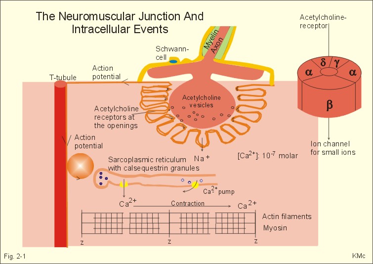
Diagram of a Neuromuscular Junction taken from Study Guide

Note that this diagram shows a neuromuscular junction of one motor neuron

The Neuromuscular Junction

diseases at the neuromuscular junction. Upper panel | Schematic diagram

between the nerve and muscle cell B. Neuromuscular junction 1.

Neuromuscular Junction · Neurons and Synapses · Synapse - Details

This gap is called the neuromuscular junction (see diagram below).

Basic structure of the neuromuscular junction showing the major channels and

affected the neuromuscular junction. Complete the following flow diagram

2-1: The neuromuscular junction and intracellular events.

in the neuromuscular junction. Diagram showing how the nerve signals the

the sensory receptors eventually travels to a neuromuscular junction,

Highly magnified view of a neuromuscular junction (Hirsch 2007).

Structure of the neuromuscular junction. The terminal of the motoneuron is

In the vertebrate neuromuscular junctions, ATP is co-stored with

interface of the nerve and muscle - the neuromuscular junction (NMJ).

The "neuromuscular junction"

a | Electron micrograph of a Drosophila melanogaster neuromuscular junction

Repairing

Label this diagram: Page 5. Overview of Neuromuscular Junction Activity
